Scapulothoracic Bursitis
Stephen Parada, M.D., Alex Girden, BA, Jon JP Warner, MD, Sept. 1, 2013
Scapulothoracic Bursitis is a medical term for the condition which causes pain in the region of the scapula in the back of the shoulder. This condition is often associated with audible and palpable crepitation, and so it has also been named “snapping scapular syndrome”. It is believed that this condition is the consequence of bursitis (inflammation) underneath the scapula. The bursa is a normal lubricating tissue which acts to allow the scapula to glide with low friction smoothly over the ribs with normal shoulder motion (Fig. 1).
This rare condition may occur as a consequence of repetitive use of the shoulder overhead causing changes to the normal mechanics of the shoulder. The result is that the muscles which control scapular motion fatigue and the scapula losses it’s normal coordination. This can result in the upper angle of the scapula rubbing (“washboarding”) over the ribs and irritating the bursa between the scapula and the ribs. (Fig. 2)
This condition is relatively rare. A search of www.google.com under “scapulothoracic bursitis” yields 14,900 results; however, a search of www.pubmed.org, which lists published scientific articles on this topic yields 26 articles. Thus, many individuals are aware of this condition yet there is little scientific evidence for diagnosis and treatment.
Fig. 1
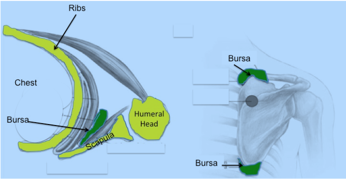
From: Conduah et al: Clinical management of scapulothoracic bursitis and the snapping scapular syndrome. Sports Health. 2010.
Fig. 2
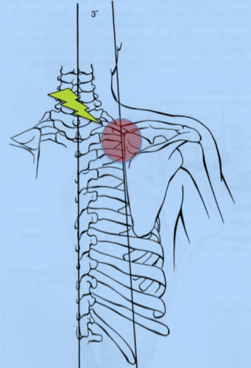
The scapula contacts the ribs at its superior (upper angle‐ red circle) and this pinches the scapulothoracic bursa creating inflammation and bursitis.
Patients often report a clicking, popping or snapping sound when they move their shoulder in a certain way, and this is usually associated with pain over the back and top of the shoulder at the upper edge of the scapula. This condition affects athletes and non‐athletes. It may occur in work activities which require repetitive overhead motion, or it may occur in sports which also repetitive overhead activities.
The examination must be performed with the shoulder and torso completely exposed so as to be able to view the shoulder girdle and the scapula position. Attention is first given to symmetry between shoulders as it is not uncommon to see postural asymmetry due to weakness of the scapular muscles. (Figs 3 & 4) (see http://www.bosshin.com/st_winging_module/)
The patient is asked to raise and lower his(her) arm and we observe motion and location of pain. Palpation (pressure) over the upper edge of the scapula usually causes discomfort.
Fig. 3
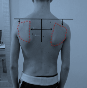
Weakness of the scapular muscles on the left result in it being lower than the right side.
Fig. 4.
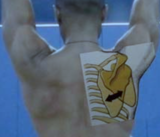
Normal motion of the scapula allows the arm to move overhead.
Regular x‐rays of the shoulder rarely demonstrate any abnormalities, though occasionally an abnormal prominent bone may be seen underneath the scapula which can cause irritation of the bursa. On occasion a CAT Scan is required to see the shape of the scapula. (Figs 5 &6)
Figure 5.
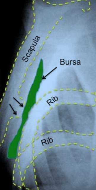
This x‐ray shows the bursa in green between the ribs and the scapula. A small growth of bone called an osteochondroma (black arrows) is compressing the bursa against the ribs.
Fig. 6.
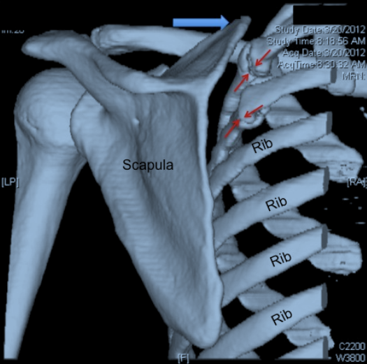
This CAT Scan has been reconstructed to show the 3‐dimensional anatomy of the scapula and the ribs. The red arrows show two unhealed rib fractures which have created inflammation and irritation of the bursa as the upper angle of the scapula (blue arrow) glides over the ribs with arm motion.
We always start with a conservative approach. The first step is to provide relief with a corticosteroid injection into the scapulothoracic bursa. This is performed in the office (Fig. 7). The injection also serves to confirm the diagnosis since if the patient derives relief the diagnosis is confirmed.
Fig. 7
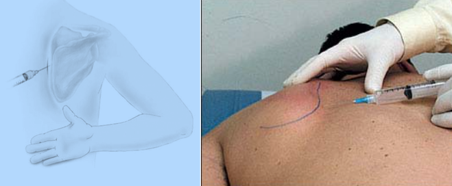
The injection is placed into the bursa just underneath the scapula.
Initial conservative treatment starts with a physical therapy program to strengthen the scapular muscles and correct any mechanical motion problems with the shoulder girdle. Anti‐inflammatory medications may also be helpful in alleviating discomfort.
Surgery is reserved only for those patients in whom pain continues to interfere with their quality of life. In our experience, response to an injection into the bursa with at least temporary relief of pain is a requirement for surgical removal of the bursa. If a patient does not derive relief from an injection it is not likely that surgery will be effective.
Removal of the scapulothoracic bursa in the case of chronic painful scapulothoracic bursitis is accomplished arthroscopically. The patient is placed on the operating table face down (prone) and the arthroscope and tissue removal instruments are inserted underneath the scapula (Fig.8 and videos).
Fig 8
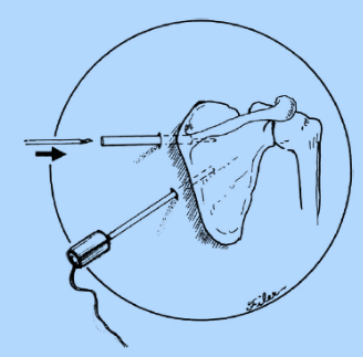
Placement of arthroscope and instruments to remove bursa.
In most cases the superior (upper) angle of the scapula bone is removed in order to eliminate the boney impingement of the scapula against the ribs which has irritated the bursa. Some surgeons may attempt to do this arthroscopically but we prefer to make a small incision and remove this bone after the arthroscopic removal of the bursa. (Figs 9 ‐ 10)
Figure 9
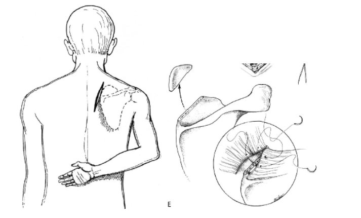
A small incision over the upper angle of the scapula allows for removal of a portion of the scapula which may be impinging the bursa between the ribs.
Fig. 10
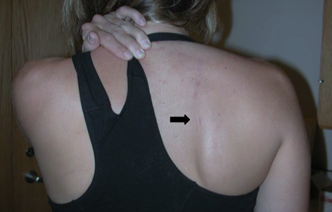
This patient had a combined open and scope procedure done, you can see the healed smaller incisions under the healed open incision after surgery.
This procedure is usually performed as a same‐day procedure so the patient goes home after surgery. The shoulder is placed in a sling for 4 weeks in order to protect the repair of the muscles to the upper scapula. Physical therapy begins after the first week as the patients begins passive motion performed by a physical therapist. After four weeks the patient can begin active range of motion for all daily living activities. After about 3 months strengthening is commenced and by 4 months all activities are permitted.
- There are numerous reports of success of surgery with this procedure:
- Blønd L, Rechter S.: Arthroscopic treatment for snapping scapula: a prospective case series. Eur J Orthop Surg Traumatol. 2013 Jan 5. [Epub ahead of print] (link this to pubmed abstract)
- Millett PJ, Gaskill TR, Horan MP, van der Meijden OA.: Technique and outcomes of arthroscopic scapulothoracic bursectomy and partial scapulectomy. Arthroscopy. 2012 Dec;28(12):1776-83
- Lehtinen JT, Macy JC, Cassinelli E, Warner JJ: The painful scapulothoracic articulation: surgical management. Clin Orthop Relat Res. 2004 Jun;(423):99-105 (link this to pubmed abstract)12 patients underwent scapulothoracic bursectomy over a five year period and after a follow-up of two years 81% of patients indicated they felt a marked improvement in their pain and function. (Fig. 11) (link this to pubmed abstract)
