Calcific Tendonitis: Diagnosis and Treatment
(Joe Eichinger, MD; Paul Yannopoulos, BA; Jon JP Warner, MD)
Calcific Tendonitis is a condition that usually affects individuals over the age of forty and is characterized by the accumulation of deposits of calcium in the rotator cuff tendons of the shoulder. The term “Calcific Tendonitis” simply means: Calcific – Calcium deposits and Tendonitis – Irritation or Inflammation of the tendon where the deposits accumulate. The reason that this problem causes shoulder pain is that calcium is an irritant in the shoulder and it causes the tendon to swell so that it pinches as it glides underneath the acromion bone of the shoulder as you raise and lower your arm.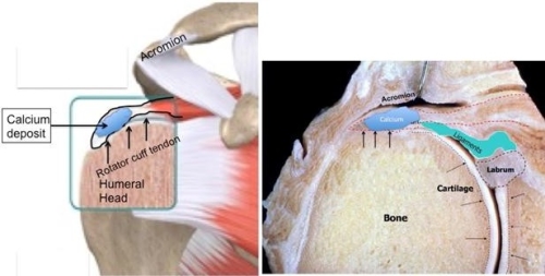

This condition usually occurs in individuals aged 30-50 years of age. Women appear to be affected more frequently than men. It is also thought that individuals with endocrine problems such as menstrual and thyroid irregularities as well as diabetes may increase the risk of developing calcific tendonitis.
The exact cause is unknown but is thought to occur because of decreased oxygen to the tendon of the rotator cuff as part of the aging process or possibly because of mechanical factors such as pressure on the tendons when the arm is lifted overhead over many years. The process of Calcific Tendonitis occurs in stages. There are generally two stages: The formative stage and the resorbtion stage. During the first stage changes in the cells of the tendon occur that allow the formation of calcium crystals. During the second stage the body reabsorbs the calcium and the tendons of the rotator cuff heal.People with calcific tendonitis may experience pain during either stage but most commonly occurs during the resorption phase. It is thought that the reason that people have pain most often during the resorption phase is because the calcium is under pressure inside the tendon.It is thought that many people (possibly up to 75% of the population) may have calcific tendonitis but it is either painless or not severe enough for them to go to the doctor.
People generally present complaining of shoulder pain. Sometimes people will lose motion and experience stiffness as a result of the calcific tendonitis. Individuals also complain of weakness and can have tenderness over the shoulder in the area where the calcium deposits occur. Simple x-rays can show calcium deposits in the rotator cuff.Other advanced imaging studies such as CT scan or MRI also show calcium deposits but are not necessary for the diagnosis.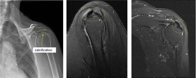

Most people will get better with conservative, non-operative treatment since the calcium deposits generally will get resorbed and go away over time. Generally, people will regain normal function of the shoulder and resolution of their pain after 2-3 weeks without any treatment. Approximately 1/3 of patients will have a complete disappearance of the calcium deposits within 3-10 years.If patients do not get better then treatment may be needed to help resolve the pain. However, some people will have persistent symptoms, principally pain. The mainstay of treatment is non-surgical and can include oral anti-inflammatory medication for pain and possibly physical therapy if stiffness or decreased range of motion is present. Another is a steroid (cortisone) injection into the shoulder. Injections and medication are useful for pain but there is no evidence that they speed the resorption of calcium. Other, less commonly used treatments include ultrasound shock therapy and “needling” in which large needles are placed into the calcium deposits to help release pressure. Ultrasound and cortisone (Iontophoresis) may be helpful in alleviating pain and resorbing calcium (See image below).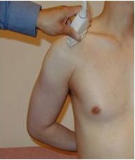

From: http://www.med.umich.edu/rad/muscskel/mskus/images/130/130.html
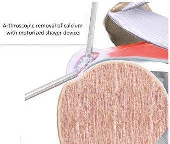
For situations in which non-surgical treatments do not help then arthroscopic surgery is used to remove the calcium deposits.
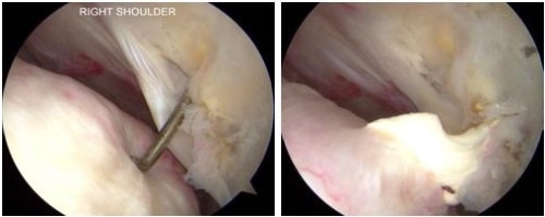
(calcium expelled from rotator cuff after arthroscopic incision. The deposit will be removed with a motorized shaving device with suction- see image which follows)
Porcellini G et al: “Arthroscopic treatment of calcifying tendonitis of the shoulderL Clinical and ultrasonographic follow-up findings at two to five years.” J Shoulder and Elbow Surg. Sept/Oct. 2004; 503-50
- Most patients improved with surgery and had minimal pain. The outcome was directly related to completeness of calcium removal
Harvie P et al: “Calcific tendonitis: Natural history and association with endocrine disorders.” J Shoulder and Elbow Surg. March/April. 2007; 169-173.
- Thyroid disorders and estrogen metabolism may increase risk of calcific tendonitis
- About one quarter of all patients develop the condition in both shoulders
- While many get better quickly about half have symptoms sufficient to require them to take time off from work.
- The majority of patients ultimately recover and the role of surgery is primarily for chronic cases of painful limitation of shoulder function
Oliva F. et al: “Calcific tendonopathy of rotator cuff tendons”. Sports Med. Arthroscopy Rev. (www.sportsmedarthro.com) Vol. 19, No 3., Sept. 2011; 237-243
- The prevalence of calcific tendinopathy is 2.7%-22% and primarily affects white women ages 30-50 years.
- 10% of the time the other shoulder is affected as well.
- In some cases the calcium deposit will resorb on its own and symptoms will resolve.
- The calcium deposit may be associated with a rotator cuff tear in 20% of cases when it is chronic.
- Non operative treatment consists of nonsteroidal anti-inflammatory drugs, cortisone injection, physical therapy to maintain motion, and modalities such as ultrasound, pulsed electromagnetic field therapy are effective in 90% of patients.
- Extracorporeal shockwave therapy (ESWT) has also been shown to be an effective treatment.
- Operative treatment is required in about 10% of patients. This is usually offered after 6 months of conservative treatment. Arthroscopic removal of calcium is a very reliable method of treatment and is successful in most patients.
