Biceps and S.L.A.P. Lesions
The Biceps muscle is a strong muscle in the front of the arm that contracts to bend the elbow.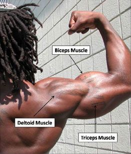
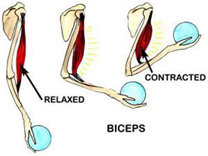
The biceps works mostly at the elbow to cause flexion of the elbow, which occurs when you do a curl as in weighlifting or when you lift anything upward toward your face.
The biceps muscle is unique because it attaches to the radius bone across the elbow and also attaches through two heads to the humerus and the scapula. Thus, it crosses both the shoulder and the elbow.
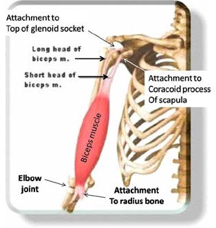
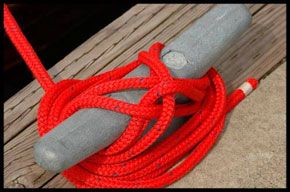
The long head of the biceps tendon runs in a groove at the top of the humerus bone and it shifts almost 90o over the humeral head before it attaches to the top of the glenoid socket. It attaches to the socket through the labrum, which is an anchoring point composed of cartilage. The labrum attachment is analogous to a cleat on a dock around which a rope (biceps) would attach.
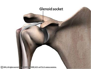
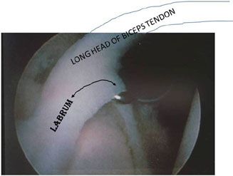
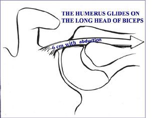
As the elbow flexes and the shoulder moves, the biceps tendon slides up and down in the groove (bicipital groove) in the front of the shoulder and may actually move up to 6 cm. Great forces are placed across the biceps with daily overhead activities such as lifting. Throwing sports place great stress on the biceps and its attachment at the superior labrum.
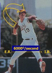
Throwing a fastball can create a velocity of arm movement of over 6000o/second and this force must be absorbed not only by the rotator cuff but also the biceps tendon and muscle as the arm is slowed to a stop after the throwing motion. This can lead to injuries to the attachment of the biceps tendon.
Biceps problems are usually either the result of a repetitive motion associated with overhead work or a sudden injury with or without a rupture of the adjacent subscapularis tendon. When the injury is a consequence of chronic inflammation, it is called biceps tendonitis. This occurs from friction and inflammation of the lubricating synovium layer that surrounds the tendon. With continued injury, the tendon may actually fray and tear the same way a rope can fray as it slides up and down repeatedly in a pulley.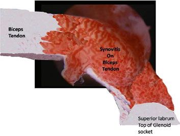
The normal biceps tendon is smooth and white. Synovitis is shown on this arthroscopic view with the magnified inflammation appearing as many small vessels over the tendon. This may be the appearance of biceps tendonitis.
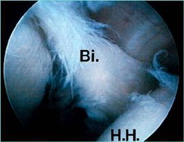
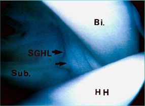
Arthroscopic view: The normal biceps tendon (Bi.) (shown on the top) is smooth and shiny. It is held in place by a ligament (SGHL) and the subscapularis tendon (Sub.). The tendon may tear from chronic wear and tear (shown on the bottom). The tendon enters the joint over the humeral head (H.H.)
In some patients, the tendon may suddenly rupture causing pain and a noticeable bulge of the biceps muscle, since the tendon retracts (pulls back) and the muscle shortens and becomes wider. This has been called a “Popeye sign” after the cartoon character who had biceps muscles with this shape.
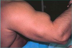

In most patients, there is no need to fix the tendon rupture because the patient may notice that the pain he/she had eventually resolves. This is due to the fact that the degenerated tendon is no longer sliding up and down in the front of the shoulder. Occasionally a patient may have pain and cramping or may not like the cosmetic deformity. In such cases a biceps tendon tenodesis is performed (described below).
In cases where the subscapularis tendon ruptures as the result of an injury, the biceps tendon may subluxate (pop out of) the groove in which it normally runs in the front of the shoulder.
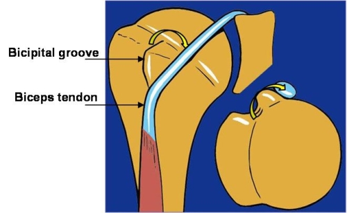
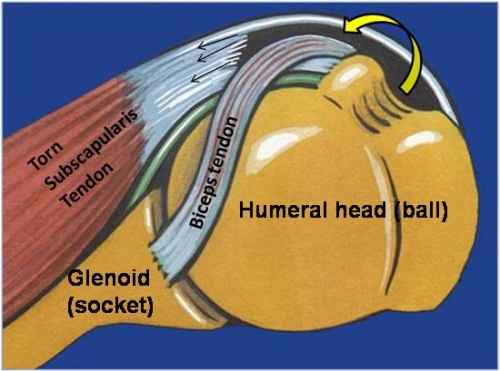
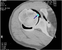
Biceps Tendon Subluxation (Courtesy Dr. Gilles Walch): The biceps tendon subluxates or pops out of the bicipital groove in the front of the humerus when the subscapularis tendon is torn. The MRI image demonstrates the tendon (arrow) out of the groove and in the joint.
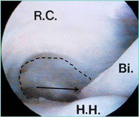
Shoulder arthroscopy demonstrates the biceps tendon (Bi.) moved out of the groove and into the joint (arrow). The position of the groove is outlined with the dotted line. The rotator cuff (R.C.) and the Humeral Head (H.H.) are labeled as well.
With biceps tendonitis, you may feel pain in the front of the shoulder. Pressure in the area of the biceps will cause pain. Use of the arm overhead may also be associated with pain in the front of the shoulder.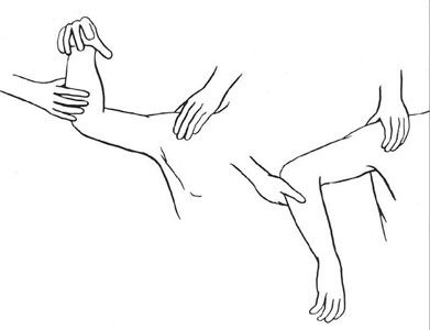
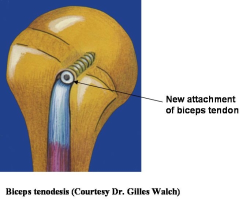
Physical therapy and activity modification may help to relieve pain. Ultrasound or Iontophoresis (ultrasound with steroid cream) may reduce inflammation. Avoidance of repetitive reaching and lifting may allow the tendon inflammation to improve. Antiinflamatory medications may also help reduce pain.Surgery is reserved only for refractory cases with marked pain and limited function. In these cases, a tenodesis of the biceps tendon is performed. This may be done as an all arthroscopic procedure or an open procedure or a combination of both. The tendon is removed from its position in the bicipital groove by detaching it from its point of attachment on the top of the socket at the superior labrum. It is then fixed into the humerus bone outside the bicipital groove so it cannot move over the shoulder joint. This procedure is usually curative of pain and it does not compromise shoulder function.
Biceps Tenodesis: Post-Operative Protocol
Biceps Tenotomy: Post-Operative Protocol
Q- If my biceps tendon ruptures, does the bulge in my arm mean I will be weaker and have pain?
A- In most cases, there is no pain with the rupture and the bulge is just a cosmetic issue. Most individuals have no pain with this. For individuals under age 50 who must do a large amount of lifting or forceful use of their arm (i.e. a carpenter, electrician, laborer) it may be advisable to have the biceps tendon fixed by performing a tenodesis in order to avoid cramping in the biceps muscle.
Q- What does the incision and cosmetics look like after a tenodesis?
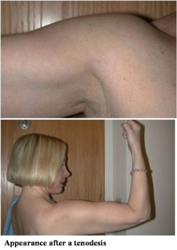
A- There are many ways to perform a tenodesis. We perform this with a small incision underneath the arm so that the tendon is retrieved underneath the pectoralis muscle and fixed to the humerus bone at this point. The tendon is first cut from its attachment in the shoulder joint while performing arthroscopy. The incision is made just underneath the arm so it is very cosmetic.
Q- How successful is a biceps tenodesis for biceps tendonitis?
A- In an analysis of over 85 patients we found that those who had the tenodesis performed by the method demonstrated above had a 98% relief of the pain they felt in the front of the shoulder.
Q- What is the recovery like and how fast can I return to active use of my arm?
A- The surgery is performed as an outpatient procedure under local anesthesia in most cases. You will be able to go home the same day. You must wear a sling for 3 weeks to protect the tendon so that it can heal in its new position on the bone. The incision should be kept dry for about one week after which it can get wet in the shower. You can use your arm to write and type, but should not actively lift with the arm. Physical therapy is usually begun after one week and active use of your arm for all daily activities can begin after 3 weeks. Strengthening of the muscle and use of the arm for sports usually takes about 3 months after surgery.
The biceps is anchored at the top edge of the glenoid socket through the labrum. The labrum is like a cleat on a dock and the biceps tendon is the rope attaching to that cleat.
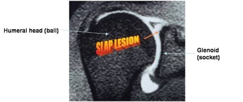
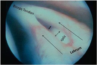
A sudden or forceful pull on the biceps as with a fall, or lifting a heavy object, or a trauma with a dislocation of the shoulder, can detach the superior labrum through a strong pull of the biceps tendon. This is termed a S.L.A.P. lesion and stands for injury to the Superior Labrum both Anterior (in front) and Posterior (behind) the biceps tendon attachment.
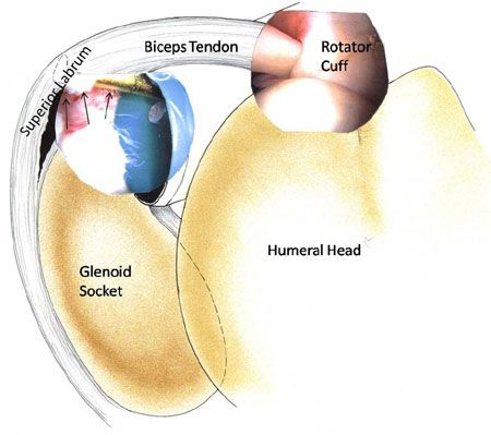
Symptoms of a S.L.A.P. Lesion: You may have pain only with use of your arm in front of your body, or you may also have audible clicking and popping in your shoulder. If you have associated instability, perhaps with a labrum tear in the front of the shoulder, you may also feel shifting in the shoulder.
Diagnosis: This injury cannot be seen on a regular x-ray. Usually an MRI-Arthrogram is required to demonstrate the S.L.A.P. lesion.
This is an artistic rendering of the view of the shoulder with arthroscopy. The superior labrum is torn and a probe inserted into the joint can lift it up away from the top of the socket (arrows). This is a typical S.L.A.P. lesion.
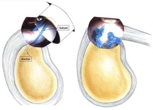
A S.L.A.P. lesion is repaired arthroscopically by placing an anchor underneath the detached labrum in the bone and then bringing the sutures from this anchor back through the labrum. These are then tied down arthroscopically compressing the labrum against the bone on the edge of the glenoid socket so that it can heal back in place.
Q- Can S.L.A.P. lesions always be repaired and should they be repaired?
A- S.L.A.P. lesions when they occur in a young athlete are usually repaired. These injuries are, however, probably less common than once thought. They may also occur in combination with an episode of instability. Sometimes the superior labrum tear is complex or it extends into the biceps tendon so it cannot be repaired. Furthermore, in individuals over age 30, it is generally advisable to perform a biceps tenodesis instead since this is associated with less pain and faster return to arm use in older individuals.
Q- When do you perform a biceps tenodesis?
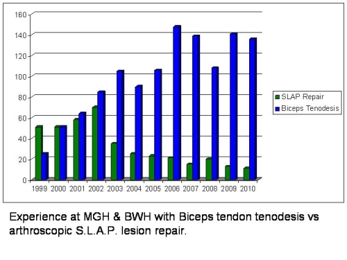
A- The biceps tendon works mainly at the elbow and only in a fast-ball pitch does it play much role in stabilizing the shoulder joint. So most individuals will not miss the tendon function at the shoulder joint if it is tenodesed as described above. Indeed, clinical experience has demonstrated that inflammation of the biceps tendon is much more common than an isolated S.L.A.P. lesion, so biceps tenodesis is performed more frequently and successfully than an arthroscopic S.L.A.P. lesion repair.

